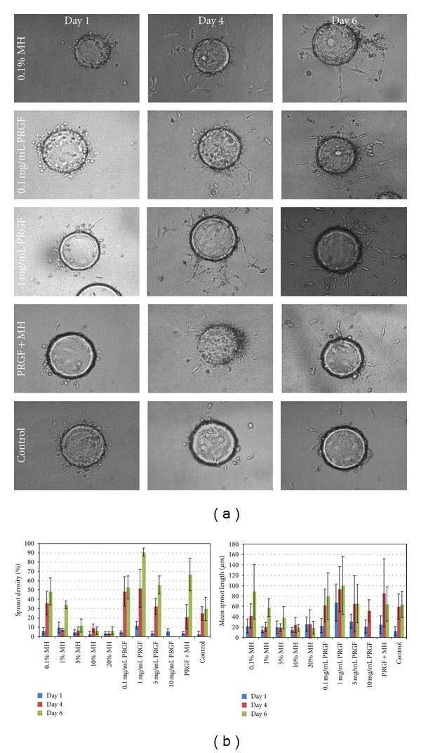
Figure 7.

Representative light microscopy images of the in vitro bead angiogenesis assay performed with hPMECs cultured on Cytodex beads. The 0.1% MH, 0.1 mg/mL PRGF, 1 mg/mL PRGF, PRGF+MH, and control are shown on days 1, 4, and 6 as those test medias resulted in maximum sprout formation (a). Mean sprout densities (b, left) and mean sprout lengths (b, right) are shown for MH, PRGF, and control medias. The results of Manuka honey supplemented media are not included as they were not significantly different from those of MH. Higher honey concentrations (5, 10, and 20% v/v) were excluded from the sprout density and sprout length graphs as they resulted in cell death.
