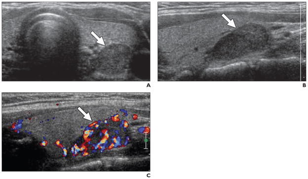
Fig. 2. 36-year-old woman with hypertension.
A, Axial gray-scale ultrasound image obtained with conventional probe shows hypoechoic lesion at left inferior thyroid bed (arrow).
B, Sagittal gray-scale ultrasound image with high-resolution probe confirms presence of lesion (arrow).
C, Lesion shows increased vascularity at color Doppler mode, consistent with parathyroid adenoma (arrow).

