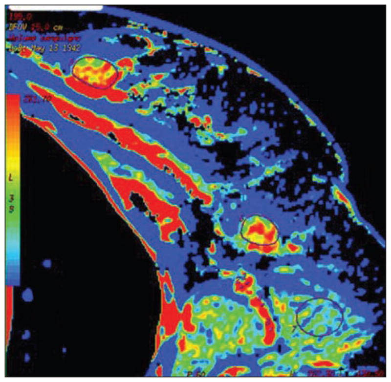
Fig. 3.

Colored map of blood volume obtained by CT perfusion in female patient with breast cancer and ipsilateral palpable node. Both primary tumor (outlined by region of interest [ROI]) and target node (outlined by ROI) are visible on this slice. High values of blood volume are depicted both in primary tumor and in target node. At postsurgical pathology, lymph node was determined to be metastatic. (Reprinted with permission from Liu Y, Bellomi M, Gatti G, Ping X. Accuracy of computed tomography perfusion in assessing metastatic involvement of enlarged axillary lymph nodes in patients with breast cancer. Breast Cancer Res 2007; 9:R40 [80])
