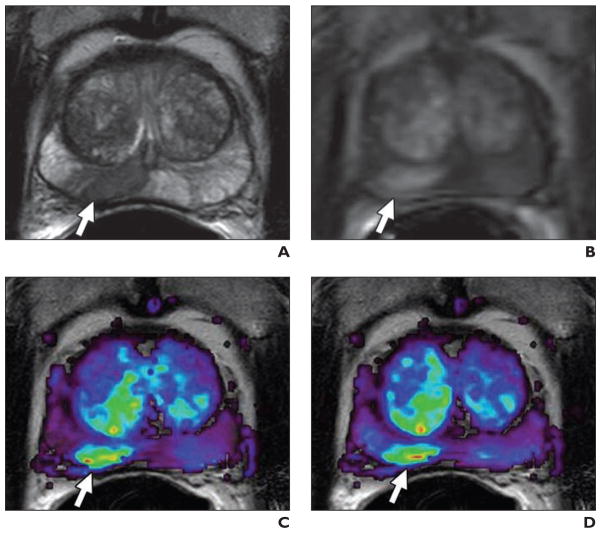
Fig. 4. 59-year-old man with prostate cancer.
A, Axial T2-weighted MR image shows low-signal-intensity foci suspicious for prostate cancer at right mid peripheral zone (arrow).
B, Raw dynamic contrast-enhanced MR image shows significant enhancement of lesion (arrow).
C and D, Corresponding Ktrans (transendothelial transport of contrast medium from vascular compartment to the tumor interstitium) (C) and kep (reverse transport parameter of contrast medium back into the vascular space) (D) maps localize right peripheral zone tumor (arrows).

