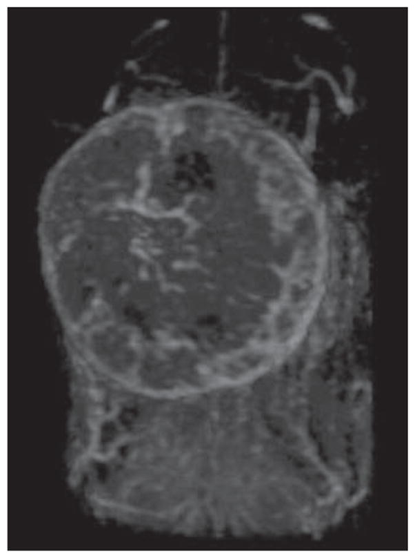
Fig. 6.

Three-dimensional dynamic contrast-enhanced MR image of orthotopically implanted breast cancer model produced with TUBO mice mammary breast cancer cell lines obtained 5 minutes after injection of 0.03 mmol Gd/kg of G6 (generation 6) dendrimer contrast agent via tail vein. Maximum-intensity- projection image cropped at site of breast tumor shows vascularity of tumor.
