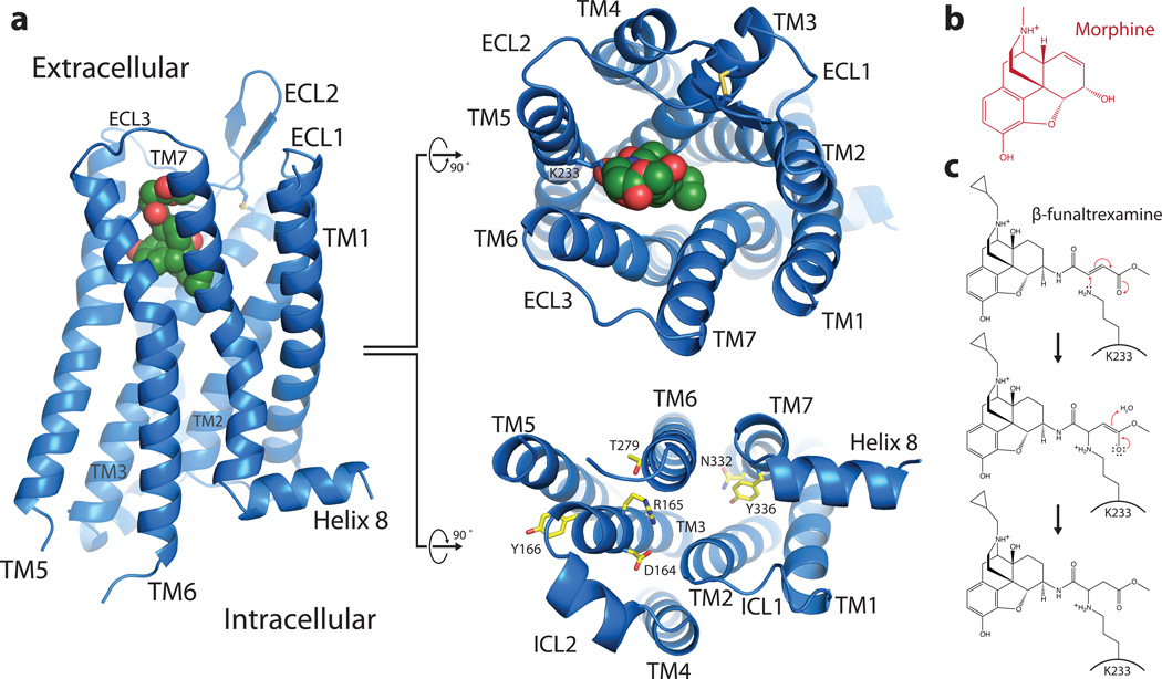
Figure 1. Overall view of μOR receptor structure.
a, Views from within the membrane plane (left), extracellular side (top, center panel) and intracellular side (bottom, center panel) show the typical seven-pass transmembrane GPCR architecture of the μOR. The ligand, β-FNA, is shown in green spheres. b, The chemical structure of morphine. c, The chemical structure of β-FNA and the chemical reaction with the side chain of K2335.39 in the receptor are shown. β-FNA is a semisynthetic opioid antagonist derived from morphine, shown at right.

