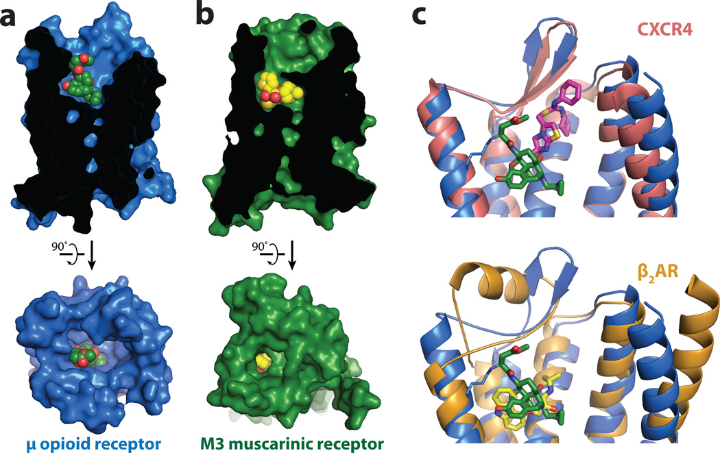
Figure 2. Comparison of ligand binding pockets.
a, The binding pocket for μOR is wide and open above the ligand, in stark contrast to the deeply buried binding pocket of the muscarinic receptors, as exemplified by the M3R shown in b. c, The small molecule antagonist IT1t (magenta) occupies a binding pocket closer to the extracellular surface of CXCR4 than β-FNA in μOR. β-FNA is positioned more similarly to the distantly related aminergic receptors as shown in c (bottom panel) for the binding site of carazolol (yellow) in the β2-adrenergic receptor (β2AR).

