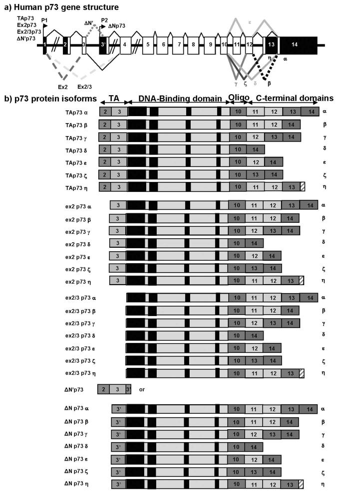
Figure 2. human p73.
a) Schema of the human p73 gene structure: Alternative splicing (α, β, γ, ζ, δ, ε, η) and alternative promoters (P1 and P2) are indicated.
b) p73 protein isoforms:
TAp73 proteins encoded from promoter P1 contain the conserved N-terminal domain (FxxψW) of transactivation (TA). Ex2p73 proteins are due to alternative splicing of exon-2. They have lost the conserved N-terminal domain (FxxψW) of transactivation (TA) but still contain part of the transactivation domain (Exon-3). Ex2/3p73 proteins are due to alternative splicing of exons 2 and 3. They have entirely lost the transactivation domain (TA) and are initiated from exon-4.
ΔN’p73 variant is often overexpressed at the mRNA level in tumours. ΔN’p73 is due to alternative splicing of exon-3′ contained in intron-3. Theoretically, ΔN’p73 mRNA would encode either for a short p73 protein or p73 protein isoforms identical to ΔNp73. ΔN’p73 mRNA contains the normal initiation site of translation in exon 2 (ATG in perfect kozak sequence) and a stop codon in exon-3′. Therefore it could encode for a short p73 protein composed only of the transactivation domain (FxxψW). It is possible that translation of ΔN’p73 mRNA is initiated from the third ATG available present in exon-3′and leading to p73 protein identical to ΔNp73 protein isoforms.
ΔNp73 proteins encoded from promoter P2 are amino-truncated proteins containing an N-terminal domain different from TAp73 proteins. Numbers indicate the exons encoding p73 protein isoforms. Black boxes indicate conserved domains.

