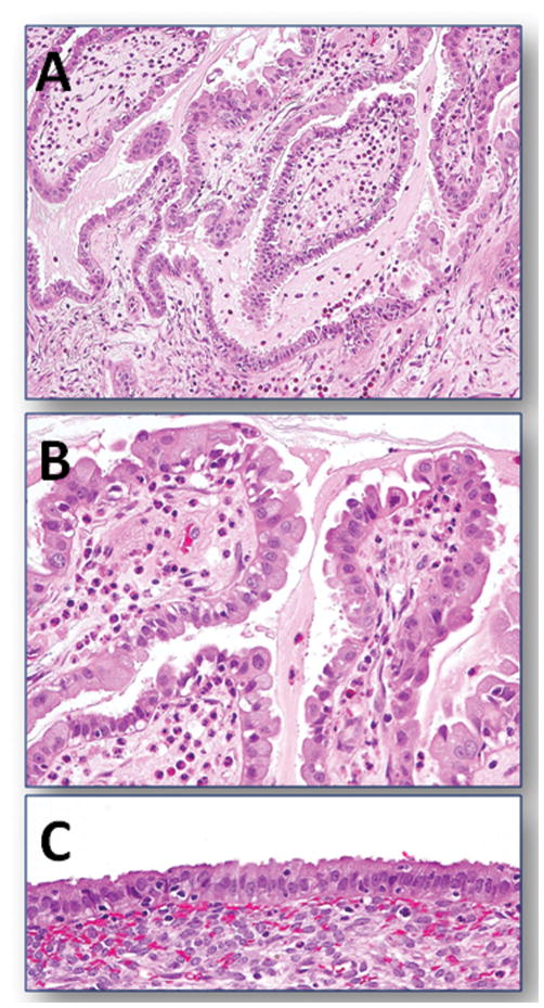
Fig. 2.
An example of a seromucinous borderline tumor of the ovary arising from an endometriotic cyst. A. A lower magnification view shows the typical papillary growth of the tumor with abundant mucinous material in the lumen. B. A higher magnification reveals the mixed histologic features of the tumor cells exhibiting serous, mucinous, and hobnail-shaped differentiation. A prominent leukocyte infiltration is also present. C. portion of endometriotic cyst adjacent to the ovarian tumor.

