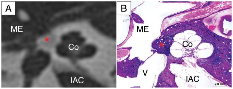
Figure 2.
The radiologic diagnosis of otosclerosis is confirmed on histology. A lucent area anterior to the oval window (*) on high resolution CT (A) matches the focus of otosclerosis (*) seen on the corresponding histologic slide, imaged at low power with light microscopy (B). Co-cochlea, ME-middle ear, IAC – internal auditory canal, V -vestibule

