Figure 3.
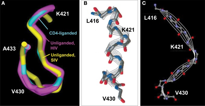
CD4BDcore conformations. (A) Superimposed CD4BDcore conformations in gp120 variants (residues 421–433) complexed with soluble CD4 and antibody 17b (cyan; 1GC1) and gp120 (extended core) without a ligand (purple; 3TGQ, 3TGR, 3TGT, 3TIH). For comparison, the CD4BDcore of unliganded SIV gp120 is included (yellow; 2BF1). Protein backbones were superimposed onto the CD4-bound structure. (B) Structure of a synthetic peptide corresponding to gp120 416–430 determined by nuclear magnetic resonance spectroscopy (NMR) in 2,2,2-trifluoroethanol. (C) Structure of the corresponding 416–430 region in the gp120 protein determined by X ray crystallography (1GC1). The NMR-derived peptide structure is reprinted with permission from reference Mihailescu et al. (2002) (Copyright 2002 American Chemical Society).

