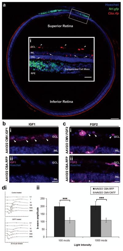
Figure 2.
In vivo investigation of neurotrophic factor expression by AAV2/2 viral vectors. Montage of confocal images (a) and a projection confocal image (a,i), from a coronal section of an adeno-associated virus vector serotype 2 chicken β-actin promoter red fluorescent protein (AAV2/2 Cba.rfp)-treated wild-type retina. (a) The control AAV2/2 CBA.RFP virus was intravitreally injected via a needle inserted through the sclera, at the inferior pars plana, and directed towards the superior retina. This resulted in the targeted transduction of inner retinal cells of the superior retina (a; red cells), at the site of cell transplantation (a,i,; Nrl.gfp; green cell mass). (a,i) The highlighted region is magnified to show the cell types transduced by the intravitreal injection of AAV2/2 viral vectors (inset i; red cells). Ganglion cells (i; white arrowheads), inner retinal neurons, and occasionally photoreceptors (i; white arrows) are transduced by the intravitreal administration of AAV2/2 viral vectors. Scale bar: 200 μm, inset 100 μm. (b) Projection confocal images of retinal sections from eyes that had been transduced with either AAV2/2 CMV.IGF1 (CMV = cytomegalovirus) (b,i) or the control AAV2/2 CBA.RFP (b,ii) viral vectors, were stained for IGF1. Increased IGF1 protein was only seen in the AAV2/2 CMV.IGF1-treated retina, at the ganglion cell layer (b,i; pink; white arrowheads). Scale bar: 20 μm. (c) Projection confocal images of retinal sections from eyes that had been transduced with either AAV2/2 CMV.FGF2 (c,i) or AAV2/2 CBA.RFP (c,ii) viral vectors were stained for FGF2. Increased FGF2 protein was observed in the GCL (c,i; pink; white arrowheads) of the AAV2/2 CMV.FGF2-treated retina only. However, nonspecific staining of the blood vessels (c,i,ii; pink; asterisks) was present in both. Scale bar: 20 μm. Nuclei were counterstained with Hoechst 33342 (blue). GCL, ganglion cell layer; INL, inner nuclear layer; ONL, outer nuclear layer; RPE, retinal pigment epithelium. (d,i) Electroretinograph traces showing a representative scotopic intensity series for both AAV2/2-treated eyes. (d,ii) Histogram demonstrating significant differences in b-wave amplitudes at both 100 and 1,000 mcds/m2 in AAV2/2 CMV.CNTF-treated eyes (mean amplitudes ± SEM; ***p < 0.001, paired t-test; N = 6).

