Figure 3.
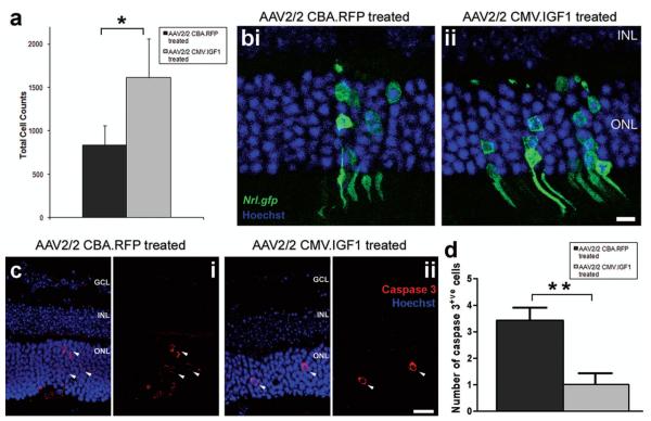
The effects of IGF1 overexpression on transplanted photoreceptor precursors and the recipient retina. (a) Histogram showing the total cell counts for AAV2/2-treated eyes. Significant enhancement of cell integration was observed for the AAV2/2 CMV.IGF1-treated eyes (mean ± SEM; *p < 0.05, paired t-test; N = 7). (b) Projection confocal images of retinal sections from eyes that have been transduced with either AAV2/2 CBA.RFP (b,i) or AAV2/2 CMV.IGF1 (b,ii), and received a subretinal cell transplantation. Integrated cells are present in both retinas (b,i,ii; Nrl.gfp; green). Scale bar: 10 μm. (c) Projection confocal images of retinal sections from eyes that have been transduced with either AAV2/2 CBA.RFP (c,i) or AAV2/2 CMV.IGF1 (c,ii), and stained for the apoptotic cell marker caspase 3 (red). Greater numbers of apoptotic cells (red; white arrowheads) were present in the control treated retina (c,i) compared to the IGF-treated retina (c,ii). Nuclei were counterstained with Hoechst 33342 (blue). GCL, ganglion cell layer; INL, inner nuclear layer; ONL, outer nuclear layer. Scale bar: 20 μm. (d) Histogram demonstrating a significant difference in the average number of caspase 3-positive cells per retinal section in the AAV2/2-treated eyes (cells/section ± SEM; **p < 0.01, paired t-test; N = 7).

