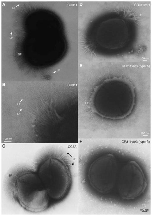
Fig. 1.
Negatively stained preparations of wild-type and fibrillar tuft variants of S. cristatus. A) Wild-type CR311 showing the organization of the short (SF) and long (LF) fibrils. B) Enlargement of the long fibrils in the fibrillar tufts of CR311. C) Wild-type CC5A showing both short and long fibrils. D) CR311var1 showing only the long fibrils that splay out at the ends. E) CR311var3 (type A) showing the remnant tuft fibrils (RF) only. F) CR311var3 (type B) having no fibrils. Magnification bar = 100 nm.

