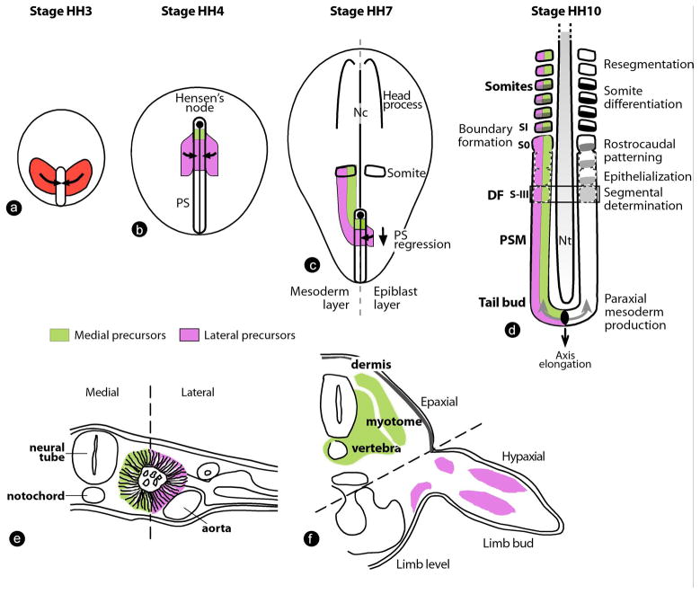
Figure 2. Paraxial mesoderm formation and segmentation in the chicken embryo.
(a–d) Dorsal views of chicken embryos. (a) Gastrulating chicken embryo at stage 3 Hamburger and Hamilton (Stage HH3) (Hamburger and Hamilton, 1992). The presumptive territory of the paraxial mesoderm (red), which contains the precursors of vertebrae and skeletal muscles, converge toward the primitive streak. (b) Stage HH4 chicken embryo. At this stage, the PS has reached its maximal length. Presumptive territories of the paraxial mesoderm are located in the superficial epiblast just below Hensen’s node (medial precursor population, green) and in two symmetrical domains located on both sides of the PS (lateral precursor population, purple). These cells are ingressing (arrows) through the PS to form the paraxial mesoderm. (c) Stage HH7 chicken embryo. The PS and node have begun their posterior regression (arrow), leaving in their wake the embryonic axis comprising the head process anteriorly and the notochord (Nc) axially. Epiblast cells (purple) continue to ingress in the PS (arrow) and join the descendents of a population of resident stem cells located in the anterior primitive streak (green) to generate the paraxial mesoderm. The mesodermal layer is represented on the left side without the superficial epiblast. (d) Posterior region of a stage HH10 chicken embryo. Somitogenesis progresses posteriorly on both sides of the neural tube (Nt) in concert with axis elongation (arrow). Paraxial mesoderm cells are produced at the tail bud level and undergo a maturation process in the presomitic mesoderm (PSM), leading to the periodic formation of new pairs of somites. Segmental determination occurs at the level of the determination front (DF, black rectangle)). Presumptive somite nomenclature according to Pourquié and Tam (Pourquie and Tam, 2001).
(e–f) Transverse sections showing the fate of medial and lateral paraxial mesoderm precursors (only the right side of the embryos are shown). The hatched line separates the medial and the lateral domains. (e) Epithelial somite in a two-day-old chicken embryo. Medial (green) and lateral (purple) somitic cells are indicated according to their origins in panels b and c. (f) Differentiation of the somitic derivatives in a five-day-old chicken embryo at the limb level. Medial somitic cells (green) contribute to epaxial body structures, such as vertebra, muscle (myotome) and dermis; whereas, lateral somitic cells (purple) give rise to hypaxial muscles in the ventral body and limbs.
PS-primitive streak; Nc-notochord; Nt-neural tube; PSM-presomitic mesoderm

