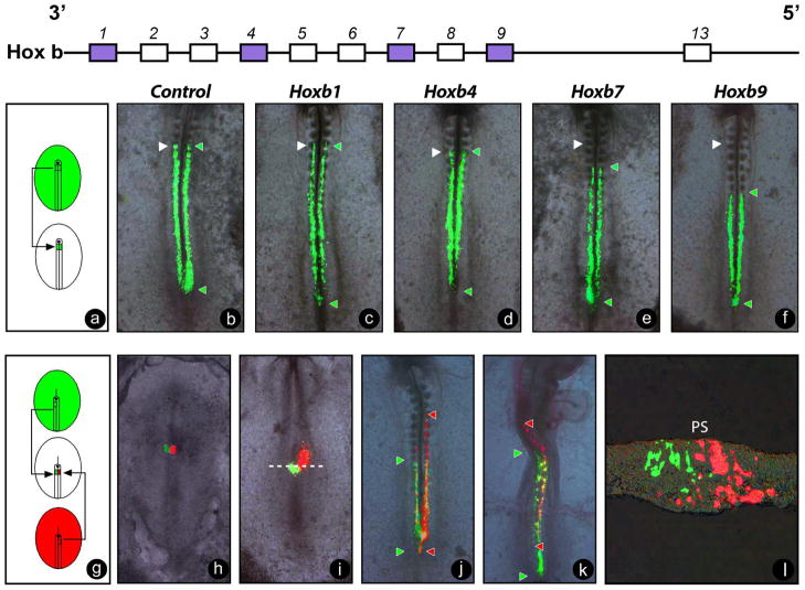
Figure 4. Hox genes control the timing of ingression of epiblast cells into the primitive streak.
(a) Schematic representation of the homotopic and homochronic grafts of fragments of the 80% level of the primitive streak electroporated with Hox-expressing constructs. (b–f) Contributions of the electroporated somitic precursors expressing control (b) or Hoxb1 (c), Hoxb4 (d), Hoxb7 (e), and Hoxb9. (f) Expression vectors driven by a ubiquitous CAGGS promoter with IRES2-ZsGreen were observed following a reincubation period of 16h. White arrowheads denote the 5th somite level, and green arrowheads denote the anterior limit of the clones overexpressing the constructs. Note the colinear distribution of the anterior limit of the green clones shifting more posteriorly as progressively more 5′ Hox genes are overexpressed.
(g) Schematic representation of homotopic and homochronic double graft of fragments of the 80% level of the primitive streak from embryos electroporated with Hoxb4-IRES2-DsRed and Hoxb9-IRES2-ZsGreen, respectively. The grafted embryo is shown just before reincubation (h), and 6h (i), 16h (j) and 40h (k) after reincubation. Green and red arrowheads denote the anterior and posterior extension of the descendants of the grafted labeled cells by each reporter along the AP axis. (h–k). The descendents of the cells expressing Hoxb4 (red) are located more anteriorly than those expressing Hoxb9 (j, k). (l) Transverse section of an embryo grafted as in (g) and incubated for 6h at the level indicated in (i) (white hatched line). Note that the cells expressing Hoxb4 (red) have entered the mesodermal layer; whereas, Hoxb9-expressiong cells (green) remain in the surface epiblast layer. Ventral views, anterior to the top. PS: primitive streak, panels h-k.

