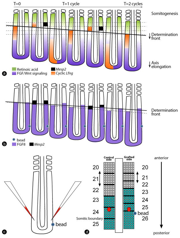
Figure 5. Interaction between Hox patterning and the segmentation machinery.
(a) The Clock and Wavefront model for somitic segment determination.
Antagonistic gradients of FGF/Wnt signaling (purple) and retinoic acid signaling (green) position the determination front (thick, black line). The periodic wave of cyclic gene expression reflecting the segmentation clock signal is shown in orange (represented on the left side only). As the embryo extends posteriorly, the determination front moves caudally. Cells that pass the determination front are exposed to the periodic clock signal, initiating the segmentation program and activating expression of gened, such as Mesp2 (black squares, represented on the right side only), in a stripe that prefigures the future segment. This establishes the segmental pattern of the presumptive somites.
(b) The graft of an Fgf8 bead in the posterior part of the embryo leads to an anterior extension of the FGF gradient (purple), corresponding to a slowing down of the determination front regression. As a result, less competent cells pass the determination front during one oscillation of the clock, leading to a smaller segment. Ultimately, this segment will form a smaller somite compared to the control side (shown in d).
(c,d) Labeling cells at the same axial position with DiI in embryos grafted with a Fgf8 bead (c) shows that cells remain at the same axial position but that the position of the somite boundaries is changed on the grafted side due to the formation of smaller somites (Dubrulle et al., 2001). (d) As a result, cells from the same axial level become incorporated into differently numbered somites. Strikingly, the expression of Hox genes (here, Hoxb9, green) is maintained at the appropriate somitic boundary rather than at the appropriate axial level, providing evidence for a cross talk between the segmentation machinery and Hox patterning. Red spots show the DiI labeled cells marked in c. Dorsal views, anterior to the top.

