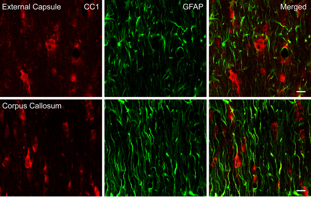
Figure 2.
Confocal micrographs of representative double-labeled CC1 and GFAP immunoreactive cells. CC1 (red) and GFAP (green) positive cells in the external capsule (Row 1) and corpus callosum (Row 2) were investigated in traumatized rats 7 days post injury. CC1 and GFAP colocalization was not observed in these regions of the rat brain (Merged). Bar = 10 microns.

