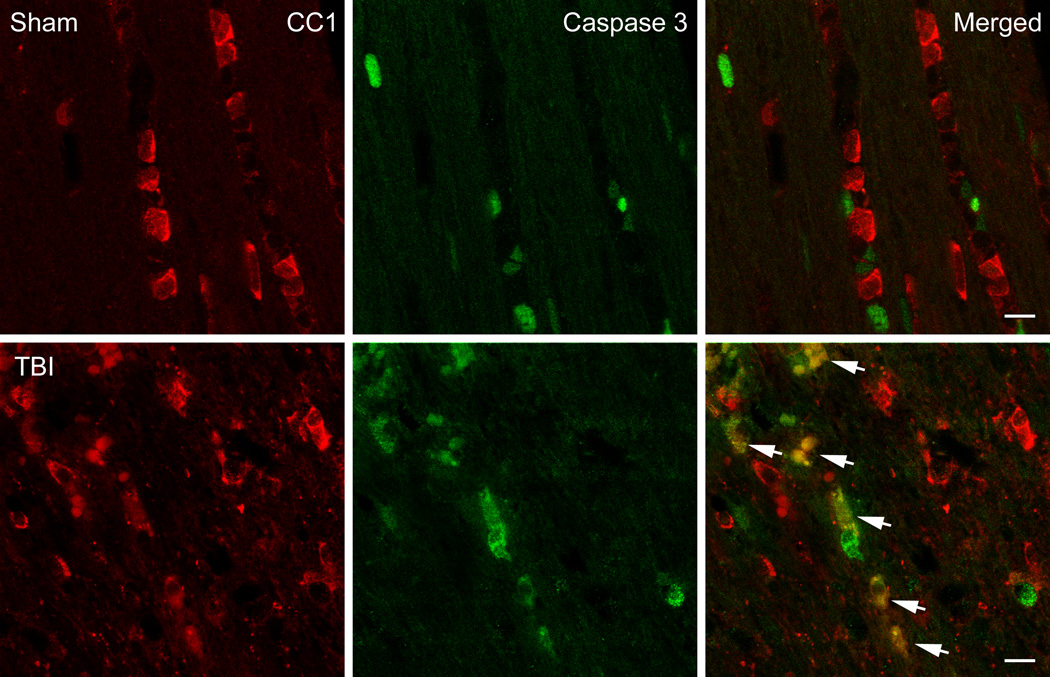
Figure 3.
Confocal micrographs of representative double-labeled CC1 (red)/Caspase 3 (green) positive cells in the external capsule. Sham animals showed CC1 positive cells with no Caspase 3 positive cells (Row 1). In contrast, a 3 day TBI animal demonstrates robust double-labeling (merged) of Caspase 3 and CC1 indicating apoptotic cell death of these oligodendrocyte (Row 2, arrows). Bar = 10 microns.

