
Figure 1. SA2-13 and its active site interactions when bound to SHV-1 S130G.
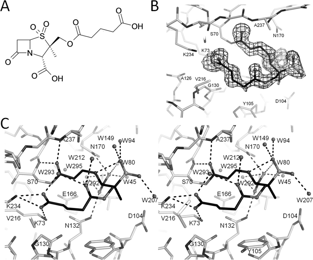
(A) Chemical structure of SA2-13. (B) Electron density of the SA2-13 compound in the active site of S130G SHV-1 β-lactamase. Unbiased omit Fo-Fc map is contoured at 2.5σ. SA2-13 carbon atoms are colored black and nitrogen, oxygen, and sulfur atoms in different shades of gray. (C) Stereo view of interactions of SA2-13 within S130G SHV-1 β-lactamase active site. Dashed black lines indicated hydrogen bonds. Water molecules are also depicted as spheres.