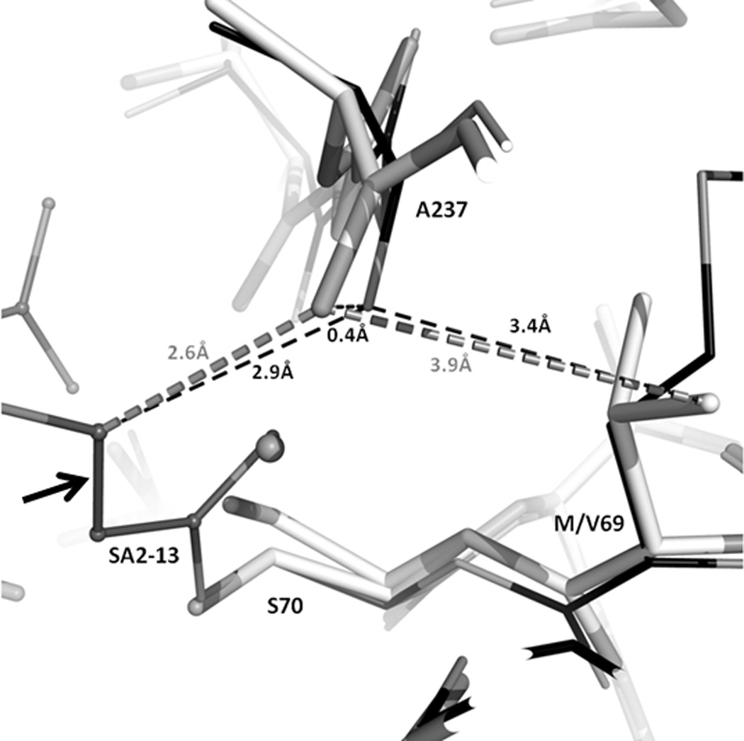
Figure 4. Comparison of M69V and wt SHV-1 structures.

Superpositioning of M69V SHV:SA2-13 (grey) with wtSHV-1:SA2-13 (black, PDB: 2H5S). Enzymes are shown as cartoon representation in the background. Dashed lines indicate distances between atoms; distances colored grey are from M69V SHV:SA2-13 and those in black are from wtSHV-1:SA2-13. Arrow indicates the trans-enamine bond of SA2-13.
