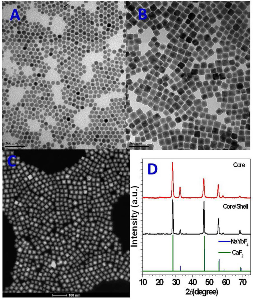
Figure 1.
Successful epitaxial growth of CaF2 shells on NaYbF4:0.5% Tm3+ core nanoparticles, resulting in uniform and monodispersed (NaYbF4:0.5% Tm3+)/CaF2 core/shell nanoparticles. Transmission electron microscopy images of a) NaYbF4:0.5% Tm3+ core and b) (NaYbF4:0.5% Tm3+)/CaF2 core/shell nanoparticles. c) High-angle annular dark-field scanning transmission electron microscopy image of (NaYbF4:0.5% Tm3+)/CaF2 nanoparticles with resolved core/shell structures; both the core (bright) and the shell (dark) are clearly visible. d) Powder x-ray diffraction patterns of NaYbF4: 0.5% Tm3+ core and (NaYbF4: 0.5% Tm3+)/CaF2 core/shell particles.

