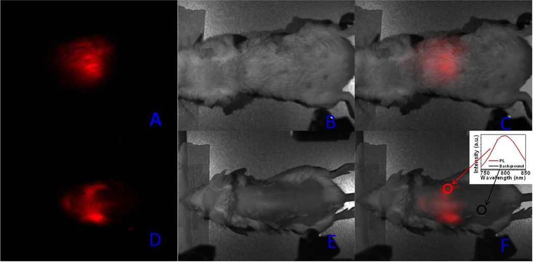
Figure 4.
Whole animal imaging of a BALB/c mouse injected via tail vein with the HA-coated α-(NaYbF4: 0.5% Tm3+)/CaF2 core/shell nanoparticles. a, d) UC PL images; b, e) bright-field images; and c, d) merged bright-field and UC PL images. Mouse was imaged in the belly (a, b, c) and the back positions. Inset in Figure 4f shows the spectra of the NIR UC PL and background taken from the circled area.

