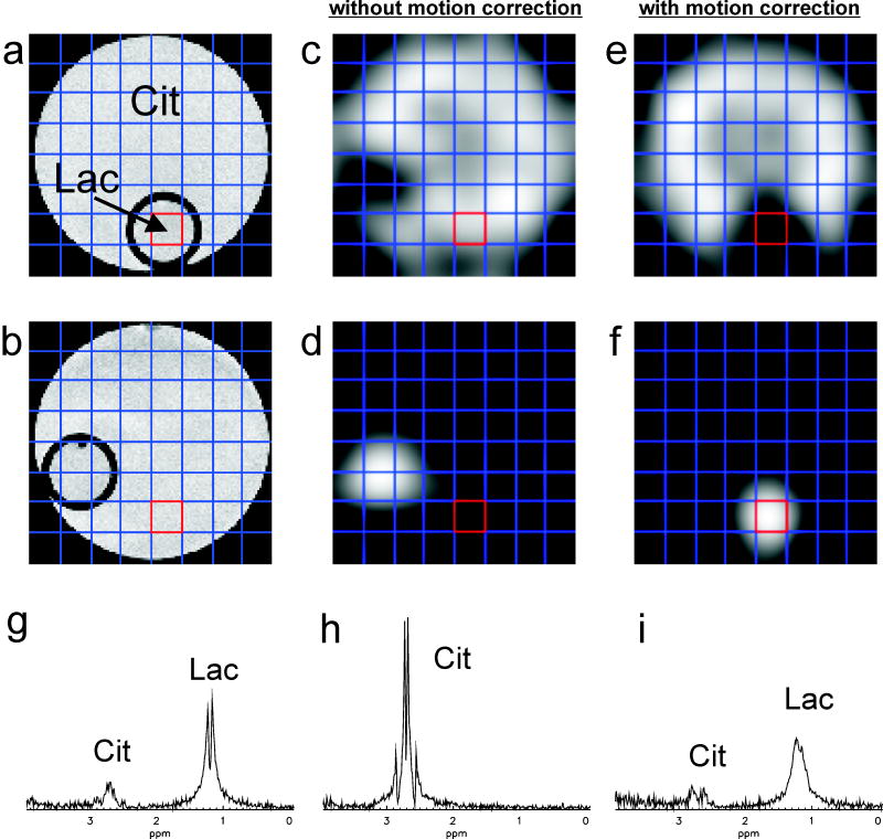
FIG. 5.
Spectroscopic imaging results for a phantom experiment with a rotation from a) to b) at the beginning of the scan: without motion correction (middle column), with motion correction (right column). Metabolite maps for Cit (c, e) and Lac (d, f) are shown for both cases. Spectra from the voxel initially situated in the compartment of the phantom containing Lac are shown for an experiment without motion (g), with uncorrected motion (h) and with corrected motion (i). The same vertical scaling was used for the representation of the three spectra.

