Figure 4.
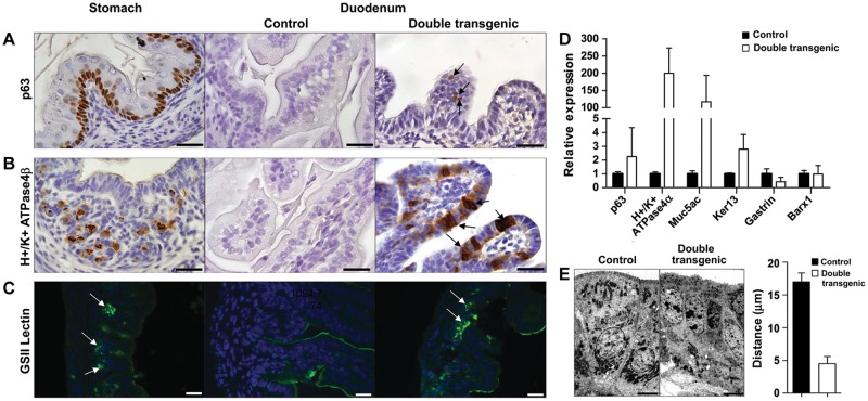
Sox2 induces stomach-like cells in the intestinal environment. Cross-sections of the stomach and duodenum at E18.5 of controls and double transgenic embryos, which received doxycycline. IHC using markers for basal cells (p63) (A) and parietal cells (H+/K+ ATPase4β) (B) showed positive staining in the stomach. Ectopically expressed Sox2 induced the appearance of basal cells and parietal cells in duodenum of double transgenic animals (arrows), whereas control duodenum is devoid of staining. Immunofluorescence for GSII lectin (C), a marker for the mucous neck cells in the stomach, showed positive cells in the double transgenic intestine (arrows), while no expression was found in the control. Scale bar, 20 μm (A–C). (D) Analysis of the expression of marker genes for basal cells (p63), suprabasal cells (Ker13), parietal cells (H+/K+ ATPase4α), and gastric pit/surface-cell mucin (Muc5ac) by qPCR showed an increase in the small intestine of double transgenic animals. No significant change in the expression level of the stomach-specific mesenchymal marker Barx1 was detected. Expression of the Gastrin hormone was reduced in double transgenic animals. (E) Nuclei of normal intestinal epithelium are oriented toward the basal membrane, whereas the nuclei of the double transgenic intestinal epithelial cells are positioned more apically, shown by EM. Bar diagram represents the quantification of the distance of the nuclei to the apical border in control and double transgenic intestines. Scale bar, 6 μm.

