Figure 3.
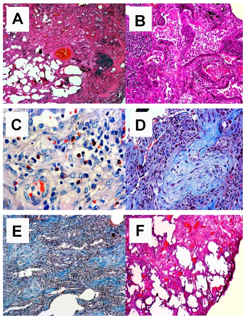
Overview of the destructive inflammatory process. (A) Acute and chronic inflammatory changes with some scarring (upper right side) are shown. HE, magnification 25×. (B) Acute alveolitis; florid inflammatory changes with highly activated pneumocytes and overspill of acute inflammatory cells into the alveoli. HE, magnification 100×. (C) Interstitial eosinophilia; in areas of longstanding inflammation one can observe a high frequency of eosinophils. They are located in the interstitium without angiocentricity and do not show overspill into the alveoli. These eosinophilic infiltrates are the major clue to a drug-induced reaction. Giemsa, magnification 200×. (D) Bronchiolitis obliterans organizing pneumonia; within the alveoli one can observe fibrobastic proliferations following acute alveolitis shown in B. CAB, magnification 100×. (E) Scarring; in more advanced stage disease one finds an interstitial accumulation of newly synthesized collagen fibers (blue color) without accompanying inflammatory infiltrate. CAB, magnification 100×. (F) End stage; interstitial scar. Note there is no honeycombing. HE, magnification 40×.
Abbreviations: HE, hematoxylin eosin; CAB, Chromotrop-Anilinblue trichrome staining method.

