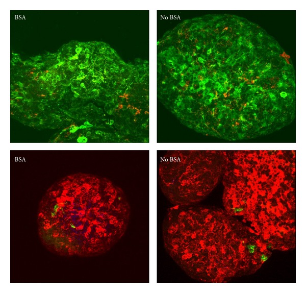
Figure 5.

Left hand panels show islets isolated with BSA while right hand panels show islets isolated without BSA. The islets were fixed in 4% PFA and cyto-spun onto glass slides before staining. Islets in top panels were stained for insulin (green) and glucagon (red). Bottom panels instead show insulin stained in red while duct cells were stained in green. Original magnification 400x.
