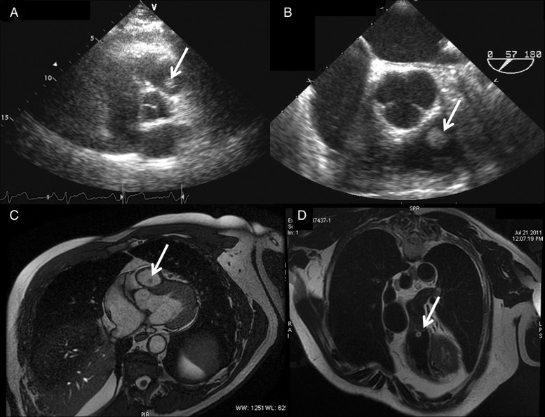
Figure 1:
(A and B) Transthoracic echocardiography and transoesophageal echocardiography showing the mass over the pulmonary valve (PV) (arrow). (C) Cardiac magnetic resonance (CMR) (Discovery 450, General Electric Healthcare, 1.5 T Unit, Milwaukee, WI, USA) scan images showing a spherical mass (arrow), 8×7.5 mm in dimension, attached to the anterior leaflet of the PV. The papillary fibroelastoma (PFE) appears hypointense compared with heart chambers in this steady-state free precession sequence. (D) CMR highlighting the PFE (arrow) which is hyperintense in black-blood T2-weighted coronal section.

