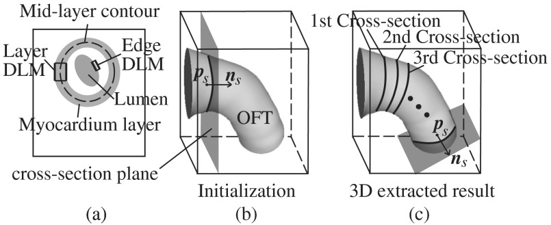
Fig. 3.
Sketch showing the OFT shape extraction procedure from OCT images. (a) Cross-sectional plane depicting the myocardium and endocardium layers, the myocardium mid-layer contour, a layer DLM placed on the myocardium and an edge DLM placed on the endocardium. (b) Initial cross-sectional plane. (c) Extraction of the cardiac shape is performed by sweeping cross-sectional planes along the OFT.

