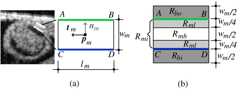
Fig. 5.
The layer double-line model (layer DLM). The layer DLM consists of two parallel lines AB (outer edge) and CD (inner edge), which can be adjusted to detect a tissue layer. (a) DLM parameters that define the DLM state : width, ; length, ; position, ; and direction, . (b) Regions of the layer DLM: the DLM is divided into an internal region, , which is in turn subdivided into two subregions, and ; and two external regions, and . These regions are used to calculate foreground and background intensity levels from OCT images.

