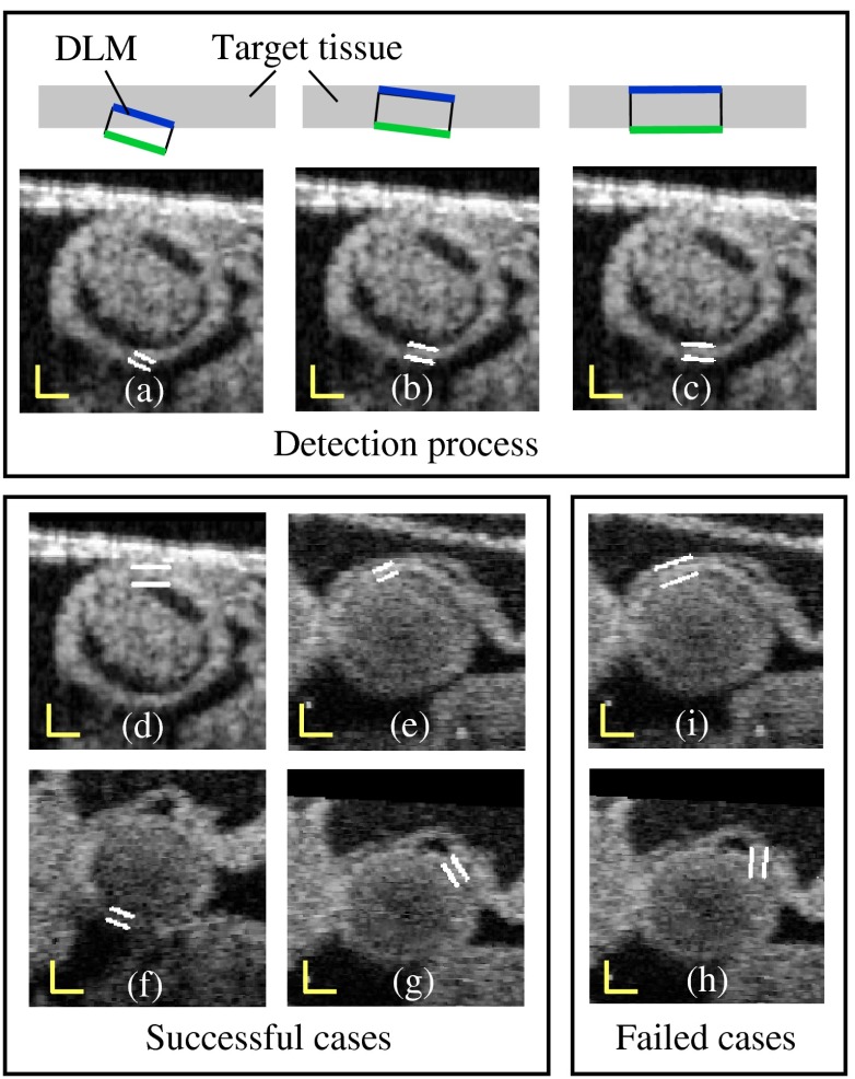
Fig. 7.
Detection of the myocardial layer (target tissue) from OCT images of the embryonic heart OFT using the layer DLM. (a–c) Detection process. (d–g) Successful segmentation results: the layer DLM can successfully detect the myocardial layer even when there are adjacent tissues (in d and e) and intensity is weak (in f and g). (h) and (i) Examples of failed detection of the myocardium: adjacent and target tissues detected together (in i) and the orientation of the DLM is incorrect (in h). Scale bars: 100 μm.

