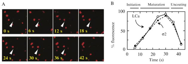
Figure 4.5.
Recruitment of clathrin and AP2 in endocytic coated pits. (A) Arrow heads point to an example of clathrin LCa-mRFP recruitment during the formation of a canonical endocytic clathrin-coated pit. (B) Plot of the fluorescence intensity normalized to the highest value before uncoating of a subset of 28 coated pits containing σ2-EGFP and LCa-mRFP, each with a lifetime of 42 s. The data were obtained using a spinning-disk confocal microscope from three BSC1 cells stably expressing σ2-EGFP and transiently expressing LCa-mRFP. (Reproduced from Fig. 4 of Ehrlich et al., 2004; from Fig. 4A and E in Ehrlich et al., 2004.)

