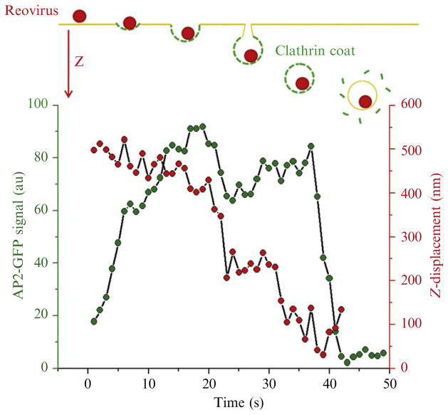
Figure 4.9.
Real-time tracking of the internalization of a reovirus particle by an endocytic AP2-containing coated carrier. Top panel: schematic representation of the experimental outcome representing the internalization of a fluorescent reovirus particle at the apical surface of a polarized MDCK cell stably expressing σ2-EGFP. Bottom panel: plot of z-displacement of reovirus and the AP2-containing pit during coated pit formation. The z-position of the virion displays an abrupt displacement toward the cytoplasm at the onset of AP2 uncoating. The data was obtained using 3D spinning-disk confocal microscopy.

