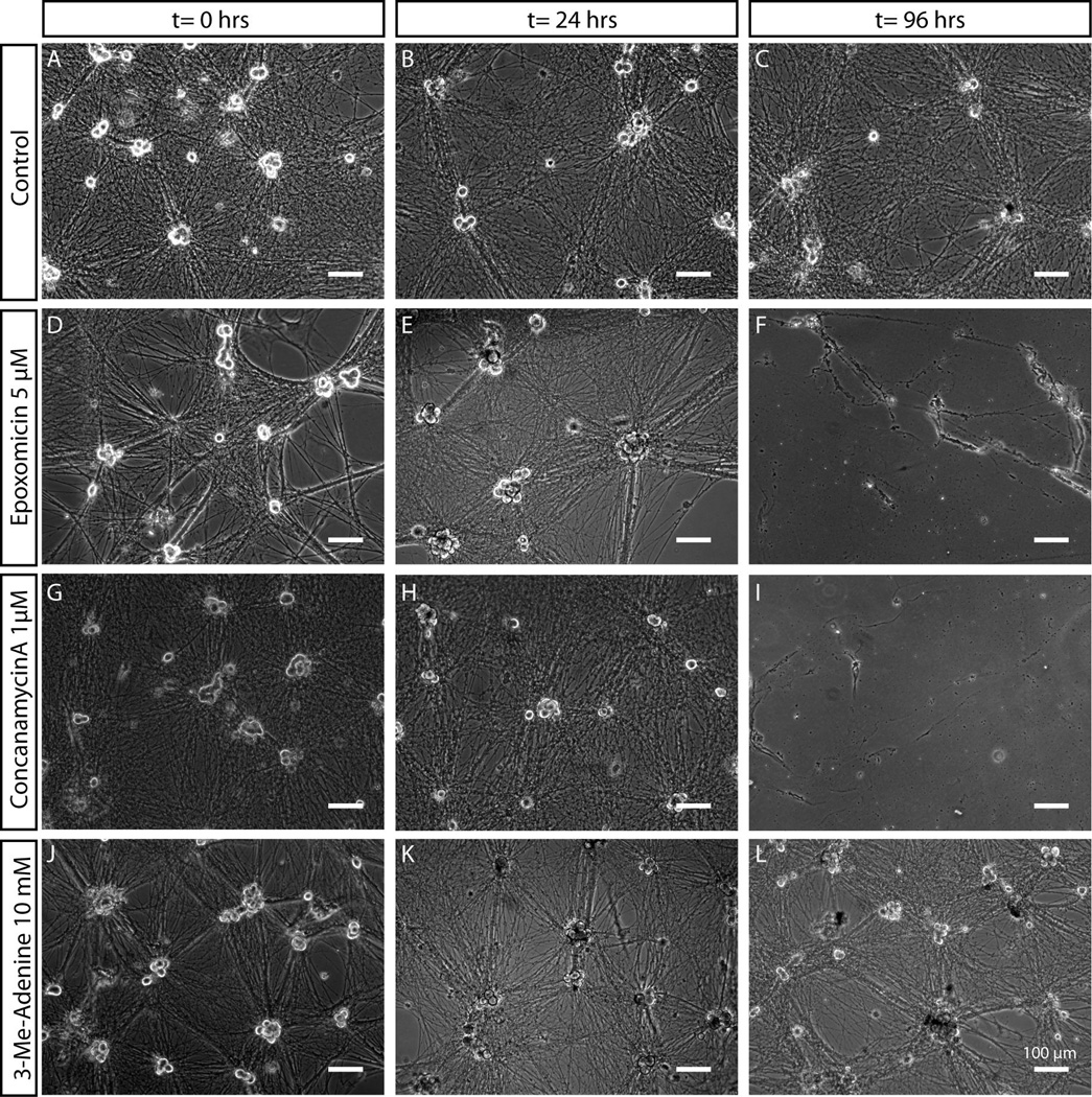
Figure 4. Differential sensitivity of sympathetic neurons to proteasome, lysosome and autophagy inhibitors.
Mass cultures of SCG neurons treated with vehicle control (A–C), epoxomicin (D–F), conconamycinA (G–I) or 3-Methyl-Adenine (J–L) were observed over the course of 96 hours. SCG morphology was unaffected upon exposure to 3-Methyl-Adenine for over 96 hours. Application of proteasome or lysosome inhibitors (epoxomicin and concanamycinA respectively), in contrast, resulted in death of SCG neurons over the course of several days, as indicated by the detachment of cell somas and presence of granulated and degenerating neuronal processes. Scale bar = 100 µm.

