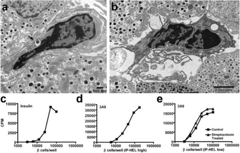
Figure 2. Islet DC present β-cell derived antigen to specific T cell hybridomas.
(a and b) Electron microscopy analysis of NOD.Rag-1-/- islets showing islet DC with an insulin granule inside a vacuole (arrow) (a) and islet DC with dendrites extending to adjacent β-cells (b). (c to e) Dispersed islet T cell assays showing: insulin-specific T cell hybridomas cultured with titrating amounts of NOD.Rag-1-/- dispersed islets (c), 3A9 T cell hybridomas cultured with dispersed islets from high producer IP-HEL mice (ILK3 strain) (d) or cultured with dispersed islets from low producer IP-HEL mice (117 strain) with or without low dose STZ treatment 4 days before islet isolation. Panel (a), (c), (d) and (e) published as Figure 3 and 6 from reference 19, with permission from PNAS. Panel (b) is from unpublished data.

