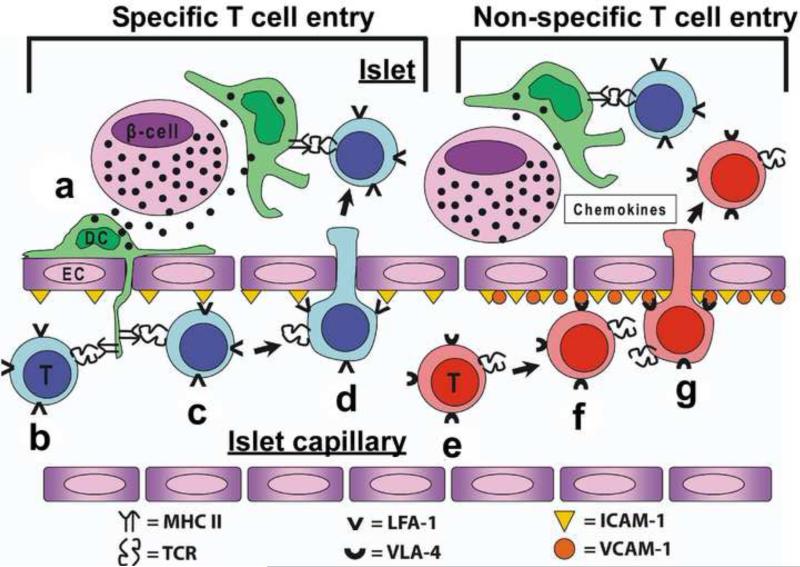
Figure 3. Proposed model of CD4 T cell entry into the islets of Langerhans Specific T cell entry.
(a) Secretory granules are taken up by the islet DC (shown in green), which are then processed and its peptides presented by MHC II. (b) Specific CD4 T cells (shown in blue) encounter their antigen presented by islet DC protruding through the fenestrated endothelium of the vessel. (c) MHC II recognition and the interaction of LFA-1/ICAM-1 favors the retention and adhesion to the endothelium of the specific T cell in the vessel. (d) The retention allows the entry of the T cell into the islet. Once inside the islet, T cell interacts with islet DC and become activated. These steps will trigger inflammatory signals and gene changes in the islet increasing ICAM-1, VCAM-1 and inflammatory chemokines. Non-specific T cell entry: (e) Non-specific T cells in circulation (shown in red) will encounter the inflammatory signals (chemokines and VCAM-1) that will allow its retention and adhesion to the islet endothelium. (f and g) Attachment to VCAM-1 and the inflammatory chemokine gradient will lead to non-specific T cell entry into the islet.

