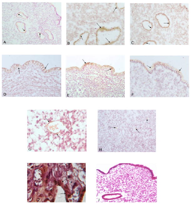
Figure 1.
Shows eNOS staining in different endometrial compartments of the study population. Panel A shows immunostaining of eNOS in glandular epithelium of a control subject, RM patient (panel B), UI patient (panel C). Panel D shows eNOS staining in luminal epithelium of a control subject (panel D), RM patient (panel E), and UI patient (panel F). Panel G shows eNOS staining in vascular endothelium, and stroma (Panel H) of a control subject. Panel I is the negative control and panel J is positive control showing eNOS staining in a human placental section. Magnification is 40x.

