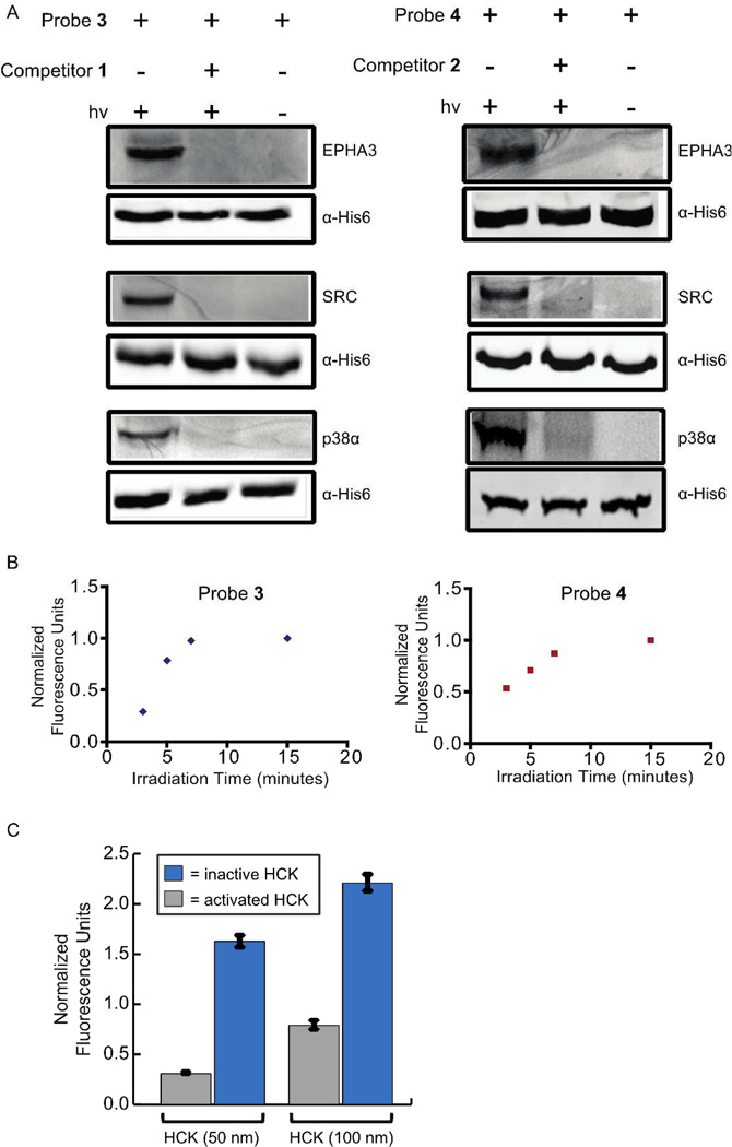
Figure 2.
Labeling of DFG-out adopting kinases with probes 3 and 4. (A) His6-tagged protein kinases (90 nM) in mammalian cell lysate (0.2 mg/mL) were incubated with probes 3 (1 µM) or 4 (1 µM) in the absence (lanes 1 and 3) or presence (lane 2) of an active site competitor (10 µM). The samples shown in lanes 1 and 2 were then irradiated with UV light for 15 minutes, while those shown in lane 3 were not. All samples were then tagged with rhodamine-azide, resolved by SDS-PAGE, and labeled proteins were detected with in-gel fluorescence scanning (top blots). Immunoblots were performed with an anti-His6 tag antibody (Cell Signaling) to ensure equal amounts of kinase were present (bottom blots). (B) The effect of UV irradiation time on photocross-linking efficiency. The tyrosine kinase SRC was incubated with probes 3 (1 µM) or 4 (1 µM) and then irradiated with 365 nm light for 3, 5, 7, or 15 minutes. Normalized fluorescence units versus irradiation time are shown. (C) Labeling of an activated and an inactive HCK construct with probe 3. Activated or inactive HCK (50 or 100 nM) in mammalian cell lysate (0.2 mg/mL) were incubated with probe 3 (1 µM). Samples were then irradiated with UV light for 15 minutes, conjugated to rhodamine-azide, resolved by SDS-PAGE, and labeled proteins were detected with in-gel fluorescence scanning. Fluorescence intensities were quantified and normalized to a fluorescent standard. The values shown are the average of assays run in triplicate ± SEM.

