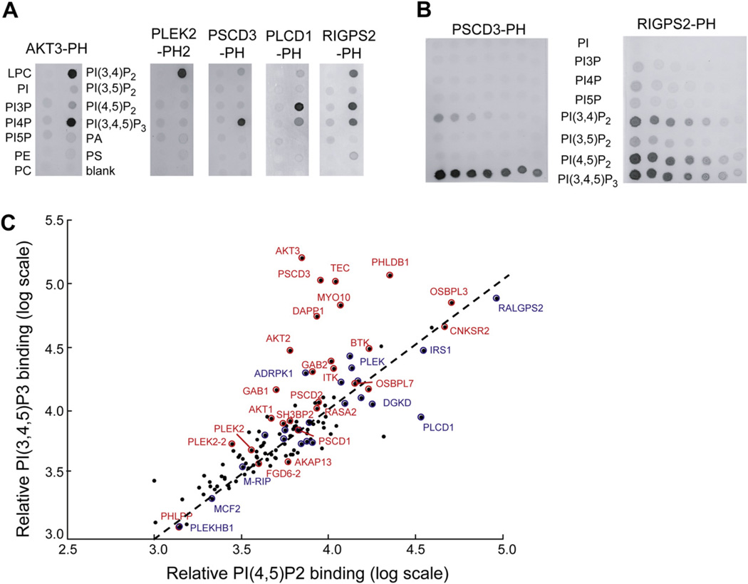
Figure 3. In Vitro Binding Selectivity of PH Domains to PI Lipids and Lipid Controls.
In vitro PI-lipid binding specificity of PH domains. Extracts of cells with overexpressed PH domains were added to PIP strips (A) and lipid arrays (B). (C) Constitutive PM targeting and receptor-triggered PM targeting partially correlate with in vitro binding to PI(4,5)P2 and PI(3,4,5)P3, respectively. PH domains with observed receptor-triggered PM translocation are shown in red. PH domains with constitutive, unregulated PM localization are shown in blue, while PH domains that remained constitutively cytosolic are marked in black without labels. A summary of all measured lipid blot binding interactions can be found in Table S2, and Figure S2 contains bar graphs showing selected lipid blot binding values.

