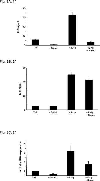
Figure 3. IL-1-mediated enhancement of secretion and expression of IL-9 is inhibited by sialoL.

(A) Naïve CD4+ T cells from BALB/c mice were stimulated (anti-CD3/CD28) under Th9-skewing conditions in the absence or presence of sialoL (3 μM) or IL-1β (10ng/ml) and a combination of both. IL-9 was determined after 72h by ELISA. (B) Naïve CD4+ T cells from BALB/c mice were stimulated (anti-CD3/CD28) under Th9-skewing conditions. After five days T cells were restimulated in the absence or presence of sialoL (3 μM) or IL-1β (10ng/ml) and a combination of both. IL-9 was determined after 48h by ELISA. (C) Naïve CD4+ T cells from BALB/c mice were stimulated (anti-CD3/CD28) under Th9-skewing conditions. After five days T cells were restimulated in the absence or presence of sialoL (3 μM) or IL-1β (10ng/ml) and a combination of both. Expression of Il9 was quantified after 48h by qRT-PCR. Similar results were obtained in four independent experiments.
