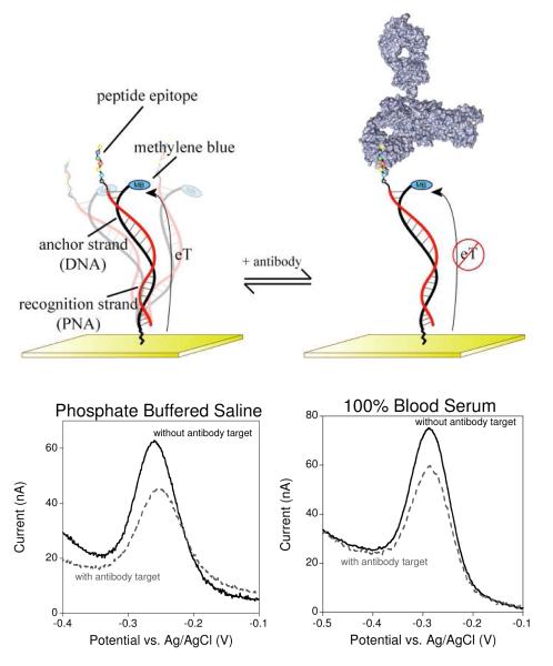
Figure 1.
The E-DNA antibody sensor (top) comprises an electrode-bound, redox-reporter-modified DNA strand, termed the “anchor strand,” that forms a duplex with a complementary “recognition strand” (here composed of PNA) to which the relevant recognition element is covalently attached. In the absence of antibody binding (top left) the flexibility of the surface attachment chemistry supports relatively efficient electron transfer between the redox reporter and the electrode surface. Binding to the relevant target antibody (top right) decreases electron transfer, presumably by reducing the efficiency with which the reporter collides with the electrode. (Bottom) Binding can thus be measured as a decrease in peak current as observed via square wave voltammetry. As shown, sensors in this class are highly selective and perform equally well in buffered saline (bottom left), undiluted blood serum (bottom right), or 1:4 diluted whole blood (see Fig. S1).

