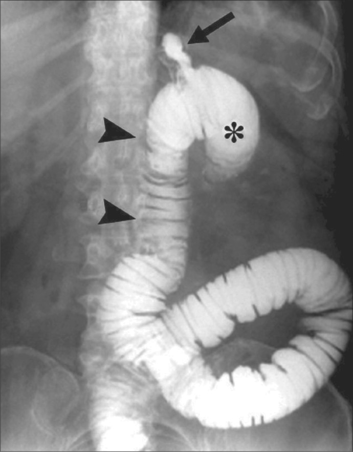
Figure 4.

Upper gastrointestinal radiograph after ingestion of contrast in a previously published patient who had had an RYGB. The upper part of the Roux limb is identified by the two arrowheads; the gastric pouch, by the arrow; and the blind limb, by the asterisk. The blind limb and the Roux limb are distended in this case because an internal hernia had caused obstruction of the jejunum. This film illustrates the ease with which a nasogastric tube could be advanced into the blind loop. Reprinted with permission from Merkle et al, 2005 (11).
