Figure 3.

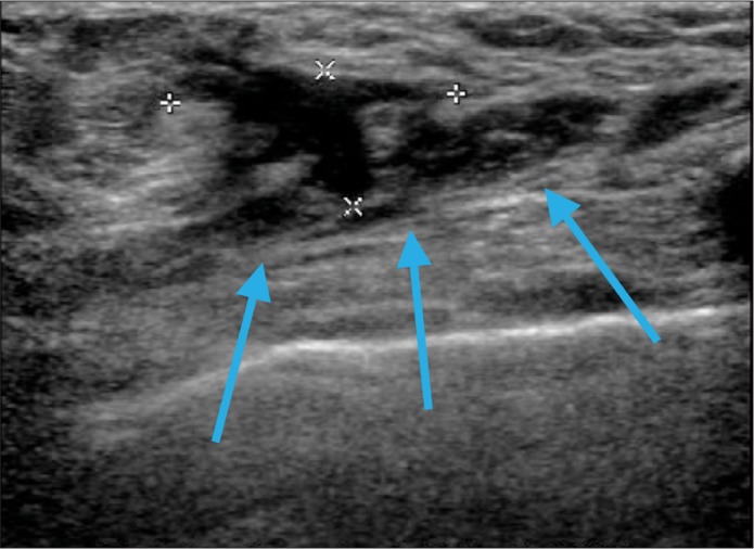
Single sonographic view of the right breast within the palpable area of concern at the 3 o'clock position, 9 cm from the nipple, demonstrates a 1.9 × 1.6 cm irregular hypoechoic lesion extending to the pectoralis muscle (arrows).

Single sonographic view of the right breast within the palpable area of concern at the 3 o'clock position, 9 cm from the nipple, demonstrates a 1.9 × 1.6 cm irregular hypoechoic lesion extending to the pectoralis muscle (arrows).