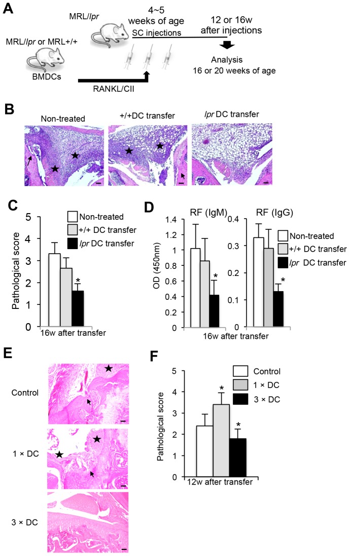
Figure 1. Therapeutic effect of repeated transfers of DCs on autoimmune arthritis.
(A) Experimental protocol is shown. BMDCs from female MRL+/+ and MRL/lpr mice were stimulated with RANKL and CII, and then female MRL/lpr mice received a total of 3 injections of the BMDCs every other day distributed over 6 day period. At 16 weeks after transfer (20 weeks of age), the recipient MRL/lpr mice were analyzed. (B) Histology of joint from recipient mice. Histological photos with HE staining are shown as representative of the recipient mice at 16 weeks after transfers. Arrow; bone erosion or synovial proliferation, star; mononuclear inflammatory infiltrate, fibrosis, or panus. Scale bar: 100 µm (n = 7, 10 and 12 per group respectively). (C) The histological score of the recipient mice was evaluated at 16 weeks after repeated transfers. Data are shown as means ± SD. (n = 7, 10 and 12 per group respectively). (D) Rheumatoid factor (RF) (IgM and IgG) antibody was measured by ELISA. Values are shown as means ± SD (n = 7, 10 and 12 per group respectively). OD = optical density. (E) RA lesions of control, a single DC transferred (1× DC), and multiple DC transferred (3× DC) MRL/lpr mice were compared. Histological photos with HE staining are shown as representative of the recipient mice at 12 weeks after transfers. Scale bar: 100 µm (n = 5 per group respectively). (F) The histological score of the recipient mice was evaluated at 12 weeks after repeated transfers. Data are shown as means ± SD (n = 5 per group respectively). *p<0.05.

