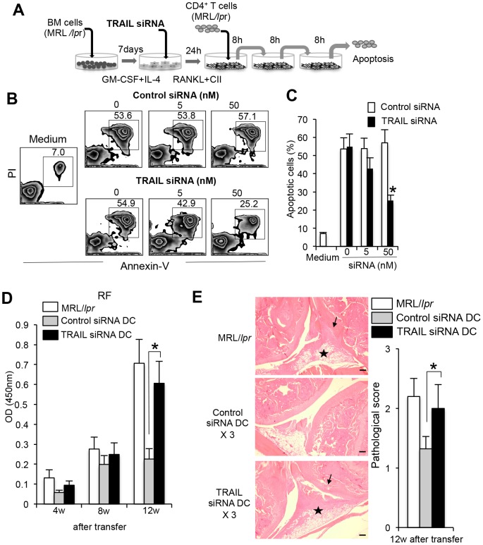
Figure 6. Regulation of Fas-independent T cell apoptosis by TRAIL siRNA-treated DCs.
(A) BMDCs from MRL/lpr mice were treated with TRAIL gene-specific siRNA or control siRNA for 24 hours, and then stimulated with RANKL and CII for 48 hours. Purified CD4+ T cells of LNs from MRL/lpr mice were repeatedly (three times) co-cultured with the activated DCs for 8 hours by transfer into each new well. (B) Expression of TRAIL on activated BMDCs treated with TRAIL siRNA or control siRNA was analyzed by flow cytometry. Results are representative of two independent experiments with similar results. (C) Apoptosis of CD4+ T cells cocultured with siRNA-treated DCs was analyzed by flow cytometry with Annexin-V and PI. Results are representative of two independent experiments. (D) Apoptotic cells (%) are shown as the mean ± SD from triplicate samples. The experiments were repeated three times with similar results. (E) In vitro TRAIL siRNA-treated and control DCs were injected three times into MRL/lpr mice (4 weeks of age). After 12 weeks after the transfers, RF level of sera from the recipient mice (16 weeks of age) was detected by ELISA. Values are means ± SD. (n = 5). The experiments were repeated twice with similar results. (F) Histology of joint from recipient mice. Histological photos with HE staining are shown as representative of five mice in each group at 12 weeks after transfers. Arrow; bone erosion or synovial proliferation, star; mononuclear inflammatory infiltrate, fibrosis, or panus. Scale bar: 100 µm. Histological score is shown as means ± SD. (n = 5) *p<0.05.

