Figure 2.
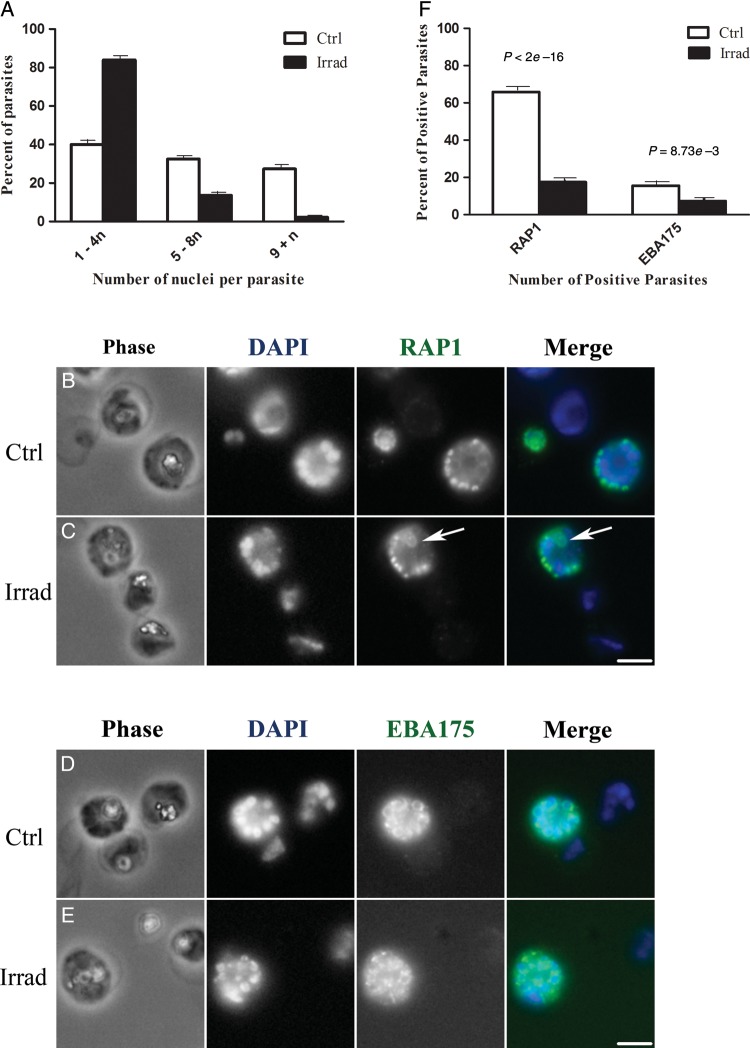
Fluorescence microscopy of purified blood-stage Plasmodium falciparum parasites from irradiated (Irrad) and control (Ctrl) cultures. A, Quantification of the number of nuclei per parasite. DAPI-stained nuclei were visually counted in approximately 600 parasites per group. In control cultures (white bars), 40%, 33%, and 27% of the parasites contained 1–4, 5–8, and ≥9 nuclei, respectively. This indicated that the parasites were developing through the entire asexual blood-stage cycle. In irradiated cultures (black bars), 84% of the parasites contained 1–4 nuclei and only 3% contained ≥9 nuclei, indicating that blood-stage parasite development was impeded. B–E, Parasite morphology is shown with phase contrast microscopy (Phase). Nuclei are labeled with DAPI (blue in merged color images), rhoptries are labeled with anti-RAP1 antibody (green in merged image), and micronemes are labeled with anti-EBA175 antibody (green in merged image). B, In control cultures, anti-RAP1 antibody stains developing rhoptries in mid-to-late schizonts. A total of 66% of control parasites (162 of 246) contain RAP1-positive vesicles that are arranged in an organized fashion around the nuclei. C, In irradiated cultures, only 18% of the parasites (56 of 319) were positive for RAP1 staining, and the RAP1 structures were sometimes clumped or had distorted morphologies (arrow). D, In control cultures, anti-EBA175 antibody stains developing micronemes in late-stage schizonts (16% of the parasites [30 of 192]), appearing as a ring around the nuclei with a brightly staining section on one side. E, In irradiated cultures, only 7% of the parasites (17 of 230) were EBA175 positive, and the stained structures were arranged in irregular patterns around the cell. F, The overall percentage of RAP1-positive and EBA 175–positive P. falciparum parasites in control and irradiated cultures. Scale bar = 4 microns.

