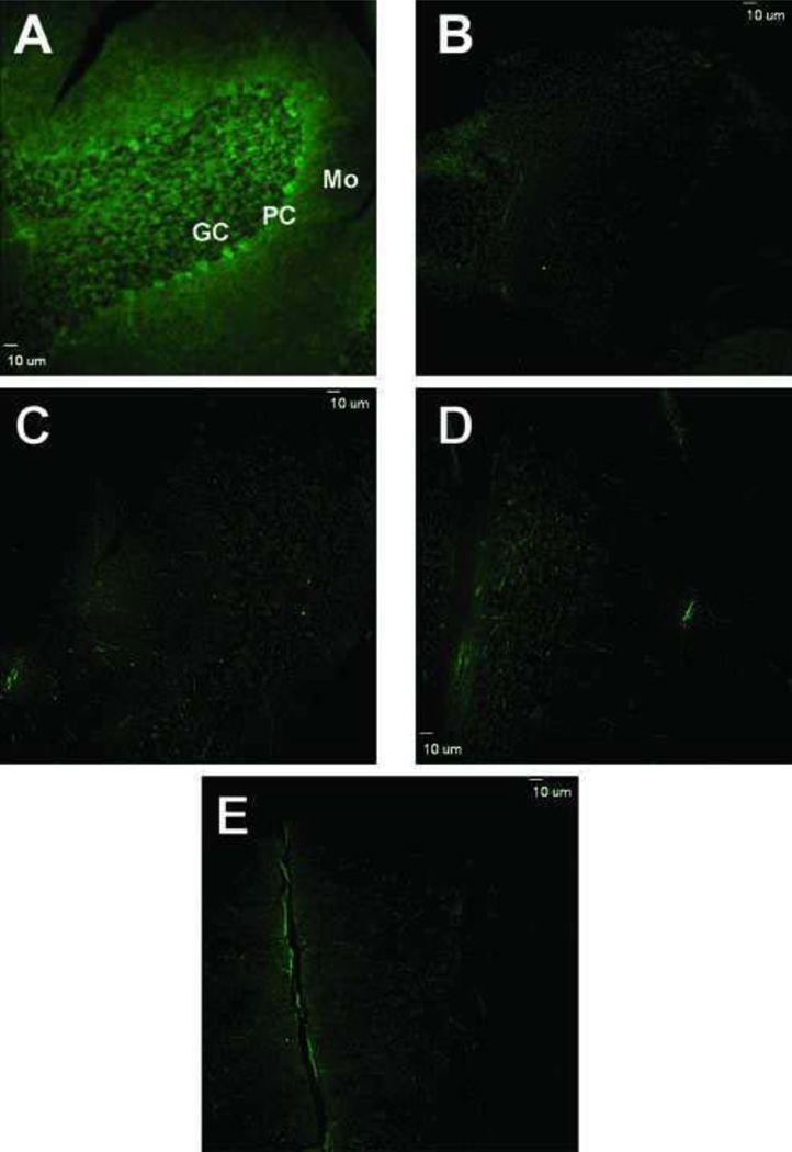
Figure 3.
Confocal fluorescence microscopy images of protein 3-NT derivatized with ABS in sections of cerebellar cortex. A) Low magnification image of cerebellar cortex that was exposed to reduction with 10 mM Na2S2O4 followed by derivatization with 2 mM ABS and 10 µM K3Fe(CN)6 as described under Methods. Controls for the specificity of labeling are shown in panels B–E where derivatization either was not performed (B) or was performed excluding ABS (C), SDT (D) or K3Fe(CN)6 (E), respectively. Bar: 10 µm.

