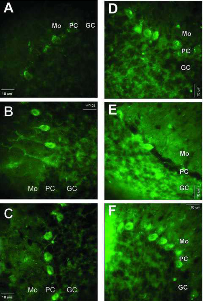
Figure 5.
Images of fluorescent labeling following either derivatization of 3-NT-containing proteins with ABS (A–C) or immunolabeling of same proteins with anti-3-NT antibody (D–F) in cerebellar cortex from slices that were pre-incubated without or with SIN-1 at varying concentrations. Sections shown in images A and D were from slices not exposed to SIN-1, whereas sections in B, C, E, and F were from slices pre-treated with either 3 mM SIN-1 (B and E) or 10 mM SIN-1 (C and F). Bar: 10 µm.

