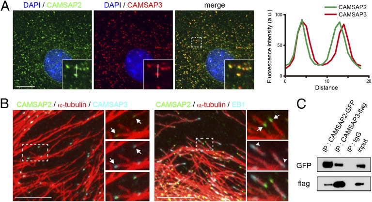
Fig. 1.
CAMSAP2 and -3 form a complex at the minus end of noncentrosomal microtubules. (A) Triple staining for CAMSAP2, CAMSAP3, and DAPI in Caco2 cells. (Inset) Enlargement of boxed area. The immunofluorescence signals of CAMSAP2 and -3 are traced along the white line, and their relative intensities are plotted at Right. (B) Triple immunostaining for CAMSAP2, CAMSAP3, and α-tubulin (Left), and for CAMSAP2, α-tubulin, and EB1 (Right). (Right) Enlarged view of boxed areas, in which the stains for CAMSAP2 (green) and CAMSAP3 (light blue) are separately shown with their merged image at the bottom. The arrows point to clusters of CAMSAP2 and -3 (Left) or CAMSAP2 (Right) attaching to an end of a microtubule. The arrowheads indicate an EB1 comet. (Scale bars, 10 μm.) (C) Coimmunoprecipitation of CAMSAP2 and -3. HEK293T cells were transiently transfected with plasmids for GFP-tagged CAMSAP2 and Flag-tagged CAMSAP3, and their lysates were subjected to immunoprecipitation (IP) with the antibodies against each tag. The precipitates were analyzed by SDS/PAGE and Western blotting using these antibodies.

