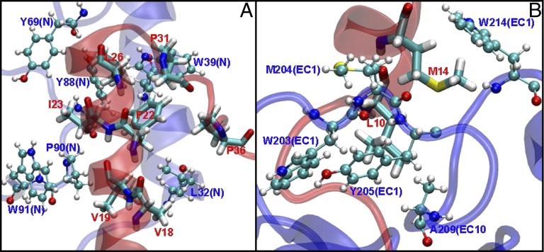
Fig. 5.
Hydrophobic interactions between GLP1R and Exe4 in the N terminus (A) and EC1 (B). Protein residues are shown in blue (CPK drawing method), whereas ligand residues are shown in red (licorice drawing method). We find 10 important hydrophobic interactions, of which 6 interactions are in the N terminus (A; all confirmed by X-ray crystal structure) and 4 interactions are in the TM bundle (B; of which 2 interactions have been confirmed by mutation studies).

